 Signs and symptoms of pulmonary
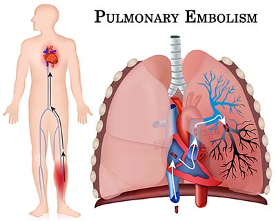
embolism depend on the size of the obstruction. If a clot obstructs a large artery, gas exchange will be severely impaired and signs and symptoms of respiratory distress will be evident. Suspect pulmonary embolism in any person with a sudden onset of unexplained dyspnea and chest pain (typically sharp and localized to a specific area of the chest) and signs of hypoxia, but who has normal breath sounds and adequate volume. The following are signs and symptoms of pulmonary embolism. However, it is important to note that the signs and symptoms of pulmonary embolism are often nonspecific and non-diagnostic. • Sudden onset of unexplained dyspnea • Signs of difficulty in breathing or respiratory distress; rapid breathing • Sudden onset of sharp, stabbing chest pain • Cough (may cough up blood) • Tachypnea • Tachycardia • Syncope (fainting) • Cool, moist skin • Restlessness, anxiety, or sense of doom • Decrease in blood pressure or hypotension (late sign) • Cyanosis (may be severe) (late sign) • Distended neck veins (late sign) • Crackles • Fever • SpO2 <94% • Signs of complete circulatory collapse In pulmonary embolism, an obstruction of blood flow in
the pulmonary arteries leads to hypoxia. Patients at risk for suffering a pulmonary embolism are those who experience long periods of immobility (such as bedridden individuals, those who travel for a long period confined in one position, those with splints to extremities) as well as those with heart disease, recent surgery, long-bone fractures, venous pooling associated with pregnancy, cancer, deep vein thrombosis (development of clots in the veins, most commonly in the legs), estrogen therapy, clotting disorders, history of previous pulmonary embolism, and those who smoke. Vascular access devices are not without problems. Since they involve a catheter inserted into the central circulation of the body, the catheter may become obstructed by clot formation at the tip. As associated emergency could be a thrombosis that forms on the catheter but then breaks off and lodges elsewhere in the body. This problem is more likely if the patient is physically inactive.
The peripheral nervous system is composed of nerves located outside the spinal cord of the brain. Afferent nerves carry sensory information from the body to the spinal cord and brain. Efferent nerves carry motor information from the brain and spinal cord to the body. Together the create a complete circuit.
Functional division of the nervous system are the voluntary nervous system and the autonomic nervous system. The voluntary nervous system includes the activity of skeletal muscles and movements. The autonomic nervous system is automatic. It influences the activity of smooth involuntary muscles and glands. It is partially independent of the rest of the nervous system. The autonomic nervous system is divided into sympathic nervous system and the parasympathetic nervous system. These two systems have opposite effects and act in a delicate balance. The sympathic nervous system is activated when the body is challenged by stressors, trauma, blood loss, fright and so on. It's known as the fright or fight response. The parasympathetic nervous returns the bodies process to normal or depresses body function. |
Archives
October 2016
Categories |
 RSS Feed